Design of Artery Phantom Prototype

Artery phantom - the artery in grey, the muscle phantom (clear) and the fluid piping connected to syringe
June 2011 - May 2012
Objective: Design and build a mechanical simulator of an artery to test the viability of using ultrasound to make blood pressure measurements.
The phantom incorporates three parts: the artery, surrounding muscle tissue, and fluid in the artery. The artery and muscle phantoms were designed to mimic the elastic stiffness of arteries and muscle tissue. The phantom was tested with static fluid under variable pressure and with fluid flow under pulsating pressure.
My responsibilities were in design conception, material background research, and final construction and testing of the assembly. The project began as undergraduate research in the Computational Instrumentation Group, MIT (PI: Dr. Brian Anthony) and documented Senior Thesis B.Sc. Mechanical Engineering
Phantom Design and Construction
Cross section of artery under Probe Force

Tissues are modeled as static springs, compressing under the downward force probe.
Clamshell Mold for Arteries

Arteries were made using cryogel. Freeze-thaw cycles control the tissue stiffness. Graphite is mixed into the cyrogel to enable opacity under ultrasound imaging. Wax molds chosen for its release properties, and milled.
Aluminum Mold for Muscle Tissue
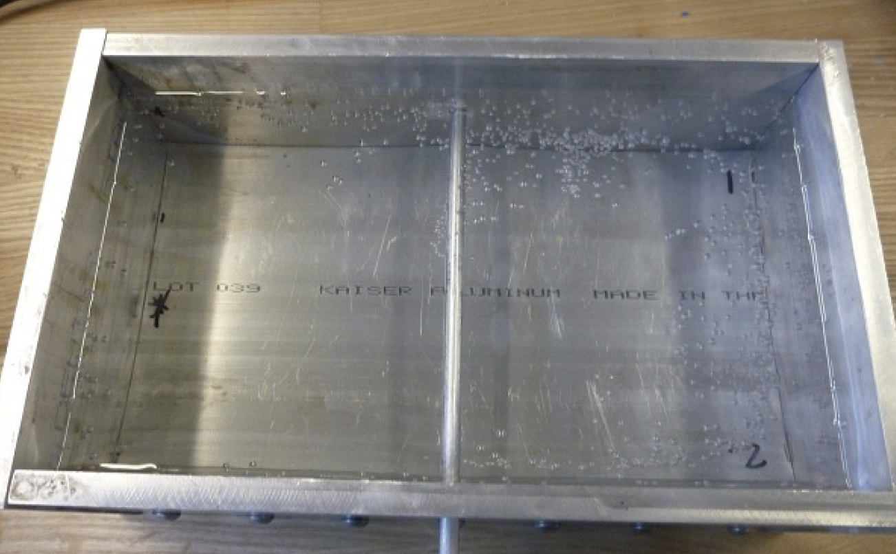
Aluminum Mold for Muscle Tissue Phantom. The phantom is made of co-polymer mineral oil-based material. It is transparent under ultrasound, enabling clear visualization of the artery phantom. The rod in the picture above provides the negative space for the artery. Copolymer concentration determines elastic stiffness.
The previous iteration constructed the phantom in two halves, but the boundary layer between the two halves was visible under ultrasound. This final iteration constructed the entire phantom as a single piece.
Static Pressure Measurement Results
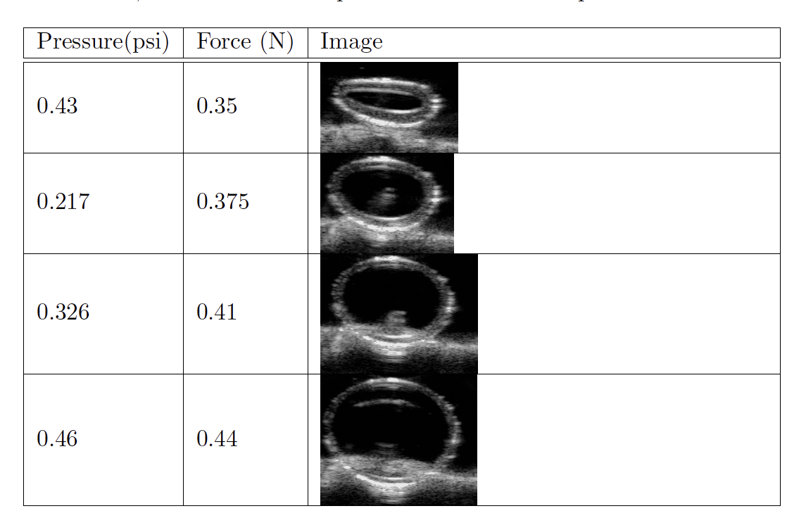
The assembly was tested with variable fluid pressure and probe forces. On left shows the ultrasound images and their accompanied force and pressure measurements. The artery wall thickness reduces under increasing fluid pressure. the vertical wall-to-wall distance responds to both probe force and fluid pressure, mimicking the static behavior of human blood vessels.
Pulsating Artery - Pump and Sensor Schematic
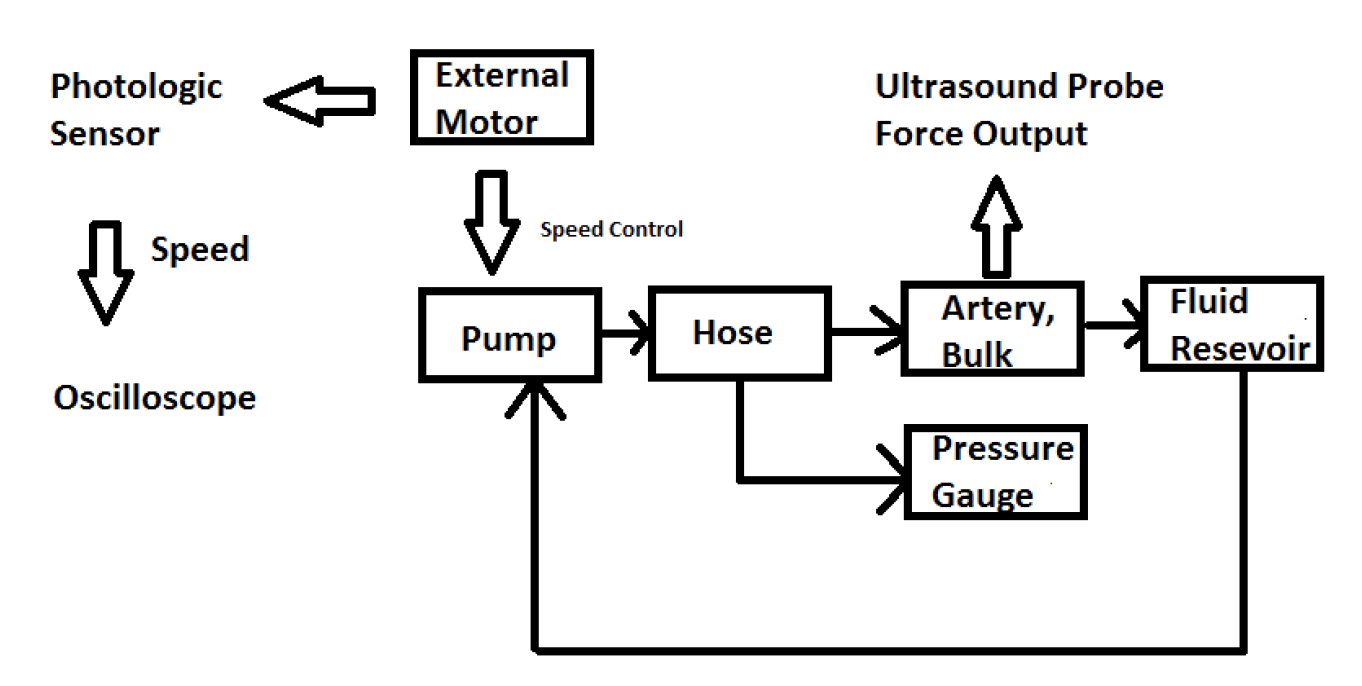

The phantom assembly was connected to a diaphragm pump to demonstrate the phantom’s ability to mimic a pulsating artery in the human body. As part of this objective, a photologic sensor was constructed and attached to the pump assembly to measure the pulse rate.
Left (top) is a schematic showing the connections amongst the phantom, pump and sensor assembly. Left (bottom) is a photograph of the assembly.
Pulsating Artery - Force Measurements at Variable Probe Compression Height
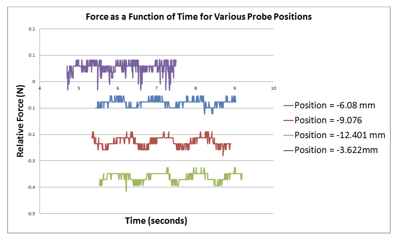
Final test of the pulsating artery was to measure the response of the artery while under variable probe compressive heights, labelled as ‘position’ in the legend. Approximate pulse is 70 beats per minute. All experimental runs shows that the force pulsates with the pulsating fluid pressure, produced by the the diaphragm pump pushing fluid through the artery. Approximate pulse rate is 70 beats/minute.